
Chronic Stress Leads to Depression: The Science Behind It
August 3, 2026


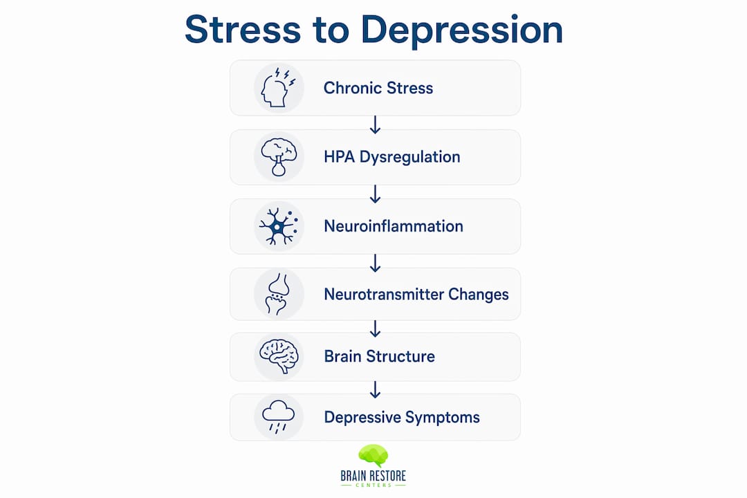
Chronic stress raises your risk for depression by triggering a cascade of biological changes: the hypothalamic-pituitary-adrenal (HPA) axis becomes dysregulated, systemic inflammation rises, monoamine neurotransmission shifts, and brain-derived neurotrophic factor (BDNF) drops, weakening the neural circuits that keep your mood stable. This is not a metaphor. These are measurable, documented changes in brain structure and immune chemistry, and they explain why prolonged stress so often crosses the line into clinical depression.
The core mechanisms work together rather than in isolation:
Population-level evidence underscores how real this risk is. During the COVID-19 pandemic, depression prevalence rose from roughly 10% to approximately 30%, a striking demonstration of what sustained social stress does at scale. The encouraging news is that these changes are largely reversible. Psychotherapy, targeted pharmacotherapy, lifestyle interventions, and integrative approaches that address inflammation and neuroplasticity can all support recovery. Understanding the mechanisms is the first step toward choosing the right path forward.
Not all stress is created equal. Acute stress, the kind you feel before a presentation or during a near-miss on the highway, is short-lived and largely adaptive. Your body mobilizes resources, you respond, and the system resets. Chronic stress is something else entirely: it is repeated or sustained activation of your stress systems over weeks, months, or longer, with no meaningful recovery window in between.
What makes chronic stress biologically distinct is not just duration. Two features amplify its impact considerably:
The clinical concept of allostatic load captures this well. Allostasis is your body’s ability to adapt to demands by changing its set points. Allostatic load is the accumulated cost of that adaptation across multiple systems, including the cardiovascular, immune, metabolic, and neuroendocrine systems. When load becomes chronic, structural brain changes accumulate in the hippocampus and prefrontal cortex rather than a simple transient stress reaction occurring and resolving. That distinction matters for treatment: you are not dealing with a mood that needs lifting, but with measurable tissue-level changes that need targeted support.

Recognizing the symptom cluster that bridges prolonged stress and a depressive episode gives you a real advantage. These signs often appear gradually, which is exactly why they are easy to dismiss as “just stress.”
Common clinical signs include:
The overlap between stress symptoms and early depression is not coincidental. Many of these signs, particularly sleep disruption and appetite changes, are direct downstream effects of the same HPA and inflammatory pathways described throughout this article. The practical distinction is persistence and functional impact: transient stress reactions typically resolve within days of the stressor easing. When symptoms persist for two or more weeks and begin affecting your work, relationships, or self-care, that is the threshold where clinical assessment becomes important.
Pro Tip: Keep a brief daily log of sleep quality, appetite, mood, and energy for two weeks. Patterns are far easier to recognize on paper than in memory, and a completed log gives a clinician far more to work with than a general “I’ve been stressed.” Most people with chronic stress develop some combination of fatigue, irritability, difficulty concentrating, appetite changes, and a perceived loss of control.

The HPA axis is your body’s primary stress-response system. Under normal conditions, the hypothalamus releases corticotropin-releasing hormone (CRH), which signals the pituitary to release adrenocorticotropic hormone (ACTH), which in turn tells the adrenal glands to produce cortisol. Cortisol then feeds back to the hypothalamus and pituitary to shut the system down. That negative feedback loop is what makes acute stress manageable.
Chronic stress breaks that loop. Repeated HPA activation leads to glucocorticoid receptor resistance, meaning cortisol can no longer effectively signal “enough.” The result is a system that either stays hyperactivated, producing chronically elevated cortisol, or eventually blunts into a state of relative hypocortisolism after prolonged dysregulation. Both patterns carry consequences.
Persistent HPA hyperactivation leads to glucocorticoid receptor resistance, which both sustains peripheral inflammation and weakens cortisol’s normal anti-inflammatory effects, contributing to elevated IL-6, IL-1β, and TNF-α and HPA axis abnormalities seen in melancholic depression.
Downstream, elevated cortisol damages hippocampal tissue, suppresses BDNF, and strips cortisol of its anti-inflammatory role, allowing immune activation to escalate unchecked. Clinically, these changes are measurable. Some studies report elevated urinary free cortisol levels in severely depressed patients compared to non-depressed groups, and the dexamethasone suppression test (DST) shows partial non-suppression in a meaningful subset of severe melancholic cases, with moderate sensitivity for that subgroup.
Pro Tip: If you are working with a clinician on stress-related mood symptoms, ask about salivary cortisol rhythm testing (morning and evening samples), urinary free cortisol, and the DST. Abnormal patterns can help distinguish HPA-driven depression from other subtypes and inform whether cortisol-modulating or anti-inflammatory strategies belong in your care plan.
Cortisol’s job, among other things, is to keep immune responses from overshooting. When glucocorticoid receptor resistance develops, that brake fails. Immune cells stop responding normally to cortisol’s suppressive signals, and pro-inflammatory cytokines including IL-6, IL-1β, and TNF-α rise in the bloodstream.
The pathway from peripheral inflammation to brain and mood involves several mechanisms:
The gut-brain axis also plays a role here. Stress-driven changes in gut permeability and microbiome composition can amplify systemic inflammation, feeding back into mood dysregulation through pathways explored in the depression-gut-hormone connection.
Up to 27% of patients with major depressive disorder show measurable neuroinflammation, and this subgroup tends to have more severe, chronic, and treatment-resistant illness. That figure matters clinically: if your depression has not responded well to standard antidepressants, inflammation may be a contributing factor worth investigating. Emerging approaches, including anti-inflammatory adjuncts and lifestyle strategies targeting immune tone, are showing promise for this subgroup.
Monoamine neurotransmitters are not simply “happiness chemicals.” They are precision signaling molecules, and chronic stress alters their synthesis, release, reuptake, and receptor sensitivity in ways that map directly onto depressive symptoms.
Here is how each system is affected:
Cytokines compound these effects by activating the enzyme indoleamine 2,3-dioxygenase (IDO), which shunts tryptophan toward the kynurenine pathway rather than serotonin synthesis. This is one reason why inflammation-linked depression often responds poorly to SSRIs alone: the problem is upstream of the reuptake transporter.
| Neurotransmitter | Stress-driven change | Depressive feature produced |
|---|---|---|
| Serotonin | Reduced synthesis and signaling | Low mood, sleep disruption, appetite changes |
| Dopamine | Depleted in reward circuits | Anhedonia, loss of motivation |
| Norepinephrine | Initial surge, then depletion | Fatigue, poor concentration, emotional blunting |
SSRIs and SNRIs address monoamine reuptake and can be effective, but their delayed onset (typically 2–6 weeks) reflects the time needed for downstream receptor and plasticity changes to accumulate. For stress-complicated or inflammation-linked depression, combining monoamine-targeted medication with anti-inflammatory strategies or neuroplasticity-focused interventions often produces better outcomes than medication alone.
BDNF is sometimes called the brain’s fertilizer: it supports the growth, maintenance, and survival of neurons, and it is critical for hippocampal neurogenesis, the process by which new neurons are generated in the adult brain. Chronic glucocorticoid exposure suppresses BDNF expression, and the hippocampus, which has a high density of glucocorticoid receptors, is particularly vulnerable.
Chronic stress biases synaptic plasticity in the hippocampus by impairing long-term potentiation (LTP) and facilitating long-term depression (LTD) in hippocampal CA1 and CA3 regions, causing dendritic atrophy and creating a brain that more readily stores and retrieves stress-related negative memories.
Human neuroimaging studies consistently show reduced hippocampal volume in people with chronic stress and major depressive disorder. Animal models fill in the cellular detail: dendritic retraction in CA3 pyramidal neurons, synapse loss, and impaired adult neurogenesis. These are not subtle changes. They translate into real functional consequences: impaired contextual memory, difficulty distinguishing past threats from present safety, and a generalized negative appraisal bias where the brain applies lessons from one stressful experience to unrelated situations.
The LTD/LTP imbalance is particularly important. LTP is the cellular mechanism underlying learning and memory consolidation. When stress facilitates LTD and impairs LTP, the brain becomes better at encoding and retrieving negative experiences and worse at forming new positive associations. That is not a psychological tendency; it is a physical change in synaptic architecture. The good news is that BDNF levels and hippocampal neurogenesis respond to exercise, sleep, and certain pharmacological interventions, meaning these changes are not permanent.
The structural and molecular changes described above do not occur in isolation. They converge on three interconnected brain regions whose coordinated function determines how you perceive, interpret, and respond to your world.
| Brain region | Effect of chronic stress | Functional consequence |
|---|---|---|
| Amygdala | Hyperreactivity and increased dendritic density | Heightened threat detection, negative emotional bias, exaggerated fear responses |
| Hippocampus | Volume loss, impaired neurogenesis, LTD facilitation | Poor contextual emotion regulation, memory generalization of stress, difficulty distinguishing safe from threatening contexts |
| Prefrontal cortex (PFC) | Dendritic retraction, weakened top-down control | Impaired cognitive control, increased rumination, reduced ability to regulate limbic responses |
The amygdala becomes a hair-trigger alarm system, flagging neutral stimuli as threatening. Normally, the hippocampus provides context (“this situation is different from the one that hurt you before”) and the ventromedial PFC applies top-down regulation (“the alarm is disproportionate; stand down”). Chronic stress weakens both of those corrective signals while amplifying the alarm.
The ventromedial PFC also projects to the ventral striatum, a key node in the brain’s reward circuitry. Reduced connectivity along this pathway maps directly onto melancholic features and anhedonia: the brain’s ability to anticipate and experience reward is structurally compromised. This circuit-level understanding helps explain why behavioral activation, a core component of cognitive behavioral therapy (CBT), works: deliberately engaging in rewarding activities helps rebuild ventral striatum activation even when motivation feels absent.
Genetics set a baseline, but early adversity can rewrite how those genes are expressed, sometimes for decades. The mechanism is epigenetic: stress-driven glucocorticoid signaling alters DNA methylation and histone modification patterns in genes that regulate the stress response itself, particularly glucocorticoid receptor genes in the hippocampus and prefrontal cortex.
Key points about how this vulnerability develops:
Childhood adversity, including abuse, neglect, household dysfunction, and early loss, is consistently associated with elevated adult inflammatory markers and higher lifetime depression risk. This is not deterministic. Protective factors, including secure attachment, social support, and early therapeutic intervention, can buffer epigenetic programming. But it does explain why two people facing the same workplace stress can have dramatically different biological and emotional outcomes.
Pro Tip: If you have a history of early adversity and are experiencing stress-related mood symptoms, tell your clinician. That history changes the biological picture: your HPA and immune systems may be more reactive than baseline measures suggest, and treatment plans that address both psychological and inflammatory components tend to work better for this profile.
Two people can face objectively similar stressors and have very different biological outcomes. The difference often comes down to perceived controllability. When a stressor feels inescapable, the brain does not signal resolution, and the HPA axis stays activated. This is the core insight from learned helplessness research: animals and humans exposed to uncontrollable stressors develop passive, withdrawn behavior that closely mirrors depression, even when escape later becomes possible.
Psychological mediators that shape the stress-to-depression pathway include:
The clinical implication is direct. Interventions that restore perceived control, such as problem-solving therapy, behavioral activation, and structured goal-setting, reduce biological stress load, not just subjective distress. Reducing rumination through mindfulness-based cognitive therapy (MBCT) or CBT techniques has measurable effects on inflammatory markers and HPA rhythm. These are not “just talking.” They are biological interventions delivered through psychological means.
There is no single moment, but there are meaningful thresholds. Allostatic load accumulates over weeks to months of sustained stress, and the transition to clinical depression typically involves both biological changes reaching a tipping point and behavioral warning signs becoming persistent enough to impair daily function.
Risk factors that accelerate the timeline include:
Population data illustrates the cumulative effect: during the COVID-19 pandemic, sustained social stress coincided with depression prevalence rising from roughly 10% to approximately 30%, a near tripling driven by the convergence of multiple risk factors simultaneously.
Warning signs that warrant clinical assessment:
If any of these apply, a clinical evaluation is the right next step, not a sign of weakness.
| Risk factor | Why it accelerates the timeline |
|---|---|
| Sleep loss | Elevates cortisol and inflammatory markers independently |
| Social isolation | Removes cortisol buffering from social contact |
| Early-life trauma | Sensitizes HPA and immune systems to adult stressors |
| Medical comorbidity | Adds to baseline inflammatory burden |
| Substance use | Disrupts HPA and neurotransmitter systems directly |
Knowing which biological pathways are driving your depression changes what treatment makes sense. A purely monoamine-focused approach may be insufficient if HPA dysregulation or neuroinflammation is the primary driver. Here is how the mechanisms map onto treatment strategies:
Pro Tip: Before your next clinical appointment, prepare three specific questions: (1) Should we test my inflammatory markers or cortisol rhythm given my stress history? (2) Is my depression profile more consistent with a monoamine, inflammatory, or neuroplasticity subtype? (3) What is the expected timeline for the approach you are recommending, and what would indicate we need to adjust?
When to investigate HPA or inflammatory markers: consider testing when depression is severe, chronic, treatment-resistant, or accompanied by significant fatigue, cognitive symptoms, or a history of early adversity. Functional medicine approaches that include inflammatory panels and cortisol rhythm testing can help personalize care, as outlined in functional medicine approaches to mental health.
| Treatment approach | Primary mechanism targeted | Expected timeline |
|---|---|---|
| CBT / behavioral activation | Psychological mediators, HPA regulation | 8 weeks |
| SSRIs / SNRIs | Monoamine signaling, downstream inflammation | 4 weeks for initial response |
| Ketamine / esketamine | Glutamate, rapid synaptic restoration | Hours to days |
| Anti-inflammatory lifestyle | Cytokine burden, HPA rhythm | Weeks to months |
| Neurofeedback / photobiomodulation | Circuit regulation, neural metabolism | Variable; adjunctive |
The mechanisms described throughout this article are not fixed. Many of the biological changes driven by chronic stress are reversible, and the most powerful tools are often the ones you can start this week.
Daily and weekly practices:
Lifestyle targets that protect neuroplasticity and reduce inflammation:
When to seek professional help:
When you do seek help, bring your symptom log, a timeline of stressors, and any relevant medical history. The more specific your account, the more targeted your clinician’s assessment can be. Resources for natural approaches to anxiety and depression can also help you prepare for that conversation.
| Step | What it targets |
|---|---|
| Consistent sleep schedule | HPA cortisol rhythm, neuroplasticity |
| Aerobic exercise 3x/week | BDNF, hippocampal neurogenesis, inflammation |
| Structured social contact | Cortisol buffering, perceived support |
| Anti-inflammatory diet | Cytokine burden, metabolic health |
| CBT / mindfulness practice | Rumination, perceived control, HPA activation |
Chronic stress leads to depression through four converging biological pathways: HPA axis dysregulation, neuroinflammation, monoamine disruption, and impaired neuroplasticity, all of which are measurable, partially reversible, and increasingly targetable with personalized treatment.
| Point | Details |
|---|---|
| HPA dysregulation is central | Glucocorticoid receptor resistance sustains cortisol dysregulation and drives downstream inflammation and brain changes. |
| Neuroinflammation affects a meaningful subgroup | Up to 27% of people with major depression show measurable neuroinflammation, often linked to treatment resistance. |
| Neurotransmitter changes are downstream, not primary | Serotonin, dopamine, and norepinephrine shifts are partly driven by cytokines and cortisol, which is why monoamine treatments alone sometimes fall short. |
| Psychological mediators amplify biology | Perceived uncontrollability and rumination extend biological stress exposure and are direct targets for psychotherapy. |
| Recovery is possible with the right approach | Exercise, sleep, psychotherapy, and inflammation-targeted strategies can reverse many stress-driven brain changes when applied consistently. |
The most important shift I see in how we approach stress-related depression at Brainrestoremeridian is moving from symptom management to mechanism identification. When someone comes in with persistent low mood, fatigue, and cognitive fog after a prolonged period of stress, the question is not just “which antidepressant?” It is: which biological pathways are most dysregulated in this person, and what combination of interventions addresses those pathways most directly?
Our assessment typically begins with a detailed stress history, not just current symptoms but the timeline, the nature of the stressors (controllable vs. inescapable), sleep patterns, and any history of early adversity. We look at functional markers where relevant, including inflammatory panels and cortisol rhythm testing, because the HPA and inflammatory picture genuinely changes what we recommend. A patient with high inflammatory markers and a history of childhood adversity needs a different care plan than someone with a recent acute stressor and no prior history.
From there, we build individualized plans that may include referral to psychotherapy (CBT or MBCT for rumination and perceived control), pharmacotherapy when indicated, and adjunctive clinic-based modalities. Neurofeedback is one tool we use to support circuit-level regulation, particularly for patients whose amygdala reactivity and prefrontal regulation are measurably impaired. QEEG brain mapping helps us see those circuit patterns directly rather than inferring them from symptoms alone. Photobiomodulation and functional medicine support are layered in when the clinical picture calls for it.
What I want readers to take away is this: the biology described in this article is not a life sentence. These pathways are responsive to intervention. The key is identifying which ones are most active in your case and building a plan that addresses them together rather than in isolation. Patient-centered decision making is at the heart of everything we do, and that means being honest about what the evidence supports, what remains emerging, and what the right referral looks like for each individual.

If you are in the Meridian, Idaho area and want to explore what a mechanism-informed assessment looks like for your situation, Brainrestoremeridian is here to help you take that next step with clarity and confidence.
These primary reviews and accessible summaries formed the evidence base for this article. Each is worth bookmarking depending on what you want to explore further.
Chronic stress, neuroinflammation, and depression: pathophysiological mechanisms and emerging anti-inflammatories (PMC): The most detailed mechanistic review available on HPA dysregulation, glucocorticoid receptor resistance, and the cytokine-depression link. Best for readers who want to understand the biological feed-forward loops in depth.
Chronic stress, neuroinflammation, and depression: an overview (Frontiers in Psychiatry): Covers the neuroinflammatory subgroup (the 27% figure), population-level evidence from the pandemic, and emerging anti-inflammatory treatment directions. Accessible for both clinical and general readers.
Central-peripheral neuroimmune dynamics in psychological stress and depression (Molecular Psychiatry): A high-level synthesis of how psychological stress activates neuroimmune and endocrine pathways, with strong coverage of epigenetic mechanisms and allostatic load. Best for readers interested in the mind-body interface.
How could stress lead to major depressive disorder? (PMC): Focuses on synaptic plasticity mechanisms, LTP/LTD imbalance, and hippocampal changes. Essential reading for understanding the memory bias and learned helplessness components of stress-induced depression.
Understanding the stress response (Harvard Health): An accessible, authoritative overview of the HPA axis and fight-or-flight response for readers who want a clear foundation before tackling the mechanistic literature.
Chronic stress: everything you need to know (Medical News Today): A patient-friendly summary of chronic stress symptoms, systemic effects, and when to seek help. Good starting point for readers early in their understanding of the topic.
Managing stress (CDC): Practical, evidence-grounded guidance on recognizing and managing long-term stress, with links to mental health resources. Useful for readers looking for structured self-management tools.
Stress (WHO): The World Health Organization’s concise overview of how stress affects mental and physical health and when it becomes a clinical concern. Best for a global public-health framing of the stress-depression relationship.
This article is general health information, not medical advice. Please consult a qualified healthcare professional for assessment and guidance specific to your situation.
Questions about your next step?
Our Meridian team can help you understand the evaluation process and which care options may fit your needs.
Schedule a ConsultationPrefer to speak with our team? Call 208-639-1397