
How Inflammation Shapes Mood Disorders and What You Can Do
August 11, 2026


Inflammation is a mechanistic contributor to a biologically distinct subset of mood disorders, and for that subgroup, it actively predicts poorer response to standard antidepressants. This is not a fringe hypothesis. A growing body of meta-analytic and translational evidence identifies elevated C-reactive protein (CRP), interleukin-6 (IL-6), and kynurenine pathway diversion as measurable, targetable features in a portion of people with depression and related mood conditions. Clinicians now refer to this cluster as the “inflamed biotype.”
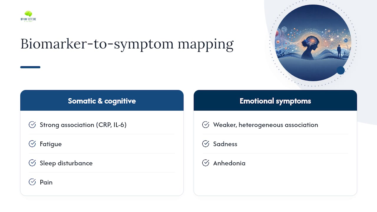
What makes this clinically meaningful is specificity. Elevated inflammatory markers are not universal in mood disorders, and they are not diagnostic on their own. They track most reliably with somatic features: fatigue, disrupted sleep, psychomotor slowing, and pain. Their association with core emotional symptoms like sadness or anhedonia is real but weaker and more variable. That distinction shapes how you interpret a lab result and how you build a treatment plan.
Inflammation does not cause depression in everyone. It causes a recognizable pattern of symptoms in a biologically identifiable subgroup, and that subgroup responds differently to treatment. Recognizing the difference is where precision psychiatry begins.
Key features of the inflamed biotype:
Inflammation contributes causally to mood disorders in a biologically identifiable subgroup, and recognizing that subgroup through biomarker testing changes both treatment selection and expected outcomes.
| Point | Details |
|---|---|
| Inflamed biotype is real | Roughly 25–45% of depressed patients show elevated inflammatory markers, with somatic symptoms and poor antidepressant response as the clearest clinical signals. |
| hs-CRP above 3 mg/L is the key threshold | This research-validated cutoff identifies patients with greater symptom severity, psychomotor slowing, and lower response to monoaminergic antidepressants. |
| Biomarker-stratified trials outperform unselected ones | Anti-inflammatory agents like celecoxib, minocycline, and infliximab show meaningful antidepressant effects specifically in patients with elevated baseline inflammation. |
| Lifestyle interventions are first-line | Aerobic exercise, Mediterranean-style diet, sleep optimization, and smoking cessation reduce CRP and IL-6 and improve mood with strong evidence. |
| Brainrestoremeridian integrates this framework | The clinic’s multidisciplinary model combines biomarker testing, lifestyle medicine, neurofeedback, and photobiomodulation to address inflammation-linked mood symptoms in Meridian, Idaho. |
Understanding the role of inflammation in mood disorders starts with a clear distinction between two related but separate processes: peripheral inflammation and neuroinflammation.
Peripheral inflammation refers to the systemic immune response circulating in the bloodstream. Its markers include cytokines such as IL-6, IL-1β, and TNF-α, along with acute-phase reactants like CRP, which the liver produces in response to cytokine signaling. This is the inflammation you can measure with a standard blood draw.
Neuroinflammation describes immune activation within the central nervous system itself. Microglia, the brain’s resident immune cells, shift from a surveillance state to an activated, pro-inflammatory state. Astrocytes become reactive. Local cytokine concentrations rise. Synaptic pruning accelerates, and neurotrophic support declines. These changes alter the chemical environment that neurons depend on for healthy signaling.
The two systems communicate through three main routes. First, systemic cytokines can cross or circumvent a compromised blood-brain barrier (BBB), which becomes more permeable under chronic inflammatory conditions. Second, the vagus nerve carries afferent immune signals directly from the periphery to brainstem nuclei, bypassing the BBB entirely. Third, cytokines reach circumventricular organs, areas of the brain with reduced BBB protection, and trigger local inflammatory cascades from there. A comprehensive PMC review details how BBB dysfunction, glymphatic impairment, and microglial recruitment together allow systemic inflammation to sustain neuroinflammation over time.
There is also a critical distinction between acute and chronic inflammation. Acute inflammation produces what researchers call “sickness behavior”: fatigue, social withdrawal, reduced appetite, and low mood. These are adaptive responses that resolve when the immune threat clears. Chronic, low-grade inflammation produces a similar but sustained pattern, and that persistence is what drives lasting mood and cognitive changes. Chronic psychosocial stress is one of the most consistent drivers of this low-grade state.
Peripheral immune activation alters brain circuits involved in mood and anxiety — a finding that has now been replicated across animal models and human neuroimaging studies, reinforcing the translational relevance of this pathway.
Common inflammatory triggers relevant to mood disorders include:
The connection between immune signaling and mood is not a single pathway. It operates through several converging molecular and cellular mechanisms, each contributing to a distinct cluster of symptoms.
When pro-inflammatory cytokines are elevated, they activate two enzymes, indoleamine 2,3-dioxygenase (IDO) and tryptophan 2,3-dioxygenase (TDO), that divert tryptophan away from serotonin synthesis and toward the kynurenine pathway. The result is a metabolic shift with two damaging consequences. Quinolinic acid, an NMDA receptor agonist with excitotoxic properties, accumulates. Kynurenic acid, a neuroprotective metabolite that normally buffers NMDA activity, decreases. This imbalance promotes glutamate-driven excitotoxicity, particularly in the hippocampus and prefrontal cortex, contributing to cognitive symptoms and impaired stress regulation. Research published in MDPI’s Cells confirms that this kynurenine pathway shift also reduces brain-derived neurotrophic factor (BDNF), directly impairing synaptic plasticity.
Cytokines reduce serotonin availability through two mechanisms: tryptophan diversion (described above) and direct suppression of serotonin transporter expression. Dopamine synthesis and release in the ventral striatum, a region central to reward processing and motivation, are also suppressed by inflammatory signaling. This is why anhedonia and anergia are so prominent in the inflamed biotype. At the glutamate level, NMDA receptor overactivity and AMPA receptor internalization alter synaptic strength in the prefrontal cortex and subgenual cingulate, regions that regulate emotional tone and rumination.
Microglial activation shifts the local brain environment toward a pro-inflammatory state. Reactive astrocytes lose their capacity to recycle glutamate efficiently, worsening excitotoxic pressure. BDNF, which supports neuronal survival, dendritic branching, and long-term potentiation, declines under sustained cytokine exposure. Mitochondrial dysfunction follows, reducing the energy supply neurons need for plasticity. The clinical correlates of these changes are anergia, psychomotor slowing, and the kind of cognitive fog that patients often describe as “thinking through mud.” Inflammation’s impact on synaptic recovery follows a similar pattern in brain injury contexts.

Chronic inflammation and HPA-axis dysregulation form a reinforcing loop. Elevated cytokines drive cortisol release, but they also induce glucocorticoid receptor resistance, meaning cortisol’s anti-inflammatory feedback signal becomes blunted. The result is sustained cortisol elevation alongside persistent inflammation, each amplifying the other. This neuroendocrine disruption contributes to sleep architecture changes, hippocampal volume loss, and impaired fear extinction.
At the network level, inflammation reduces functional connectivity between the prefrontal cortex and hippocampus, impairing top-down regulation of the amygdala. Striatal reward circuitry becomes hyporesponsive. The default mode network, already implicated in rumination, shows altered dynamics. These circuit changes map directly onto the clinical features of the inflamed biotype: reduced motivation, impaired concentration, emotional blunting, and poor stress resilience.
The pathway in summary: a peripheral inflammatory trigger raises circulating cytokines, which signal across the BBB through multiple routes, activate microglia and astrocytes, divert tryptophan toward neurotoxic kynurenine metabolites, suppress monoamine synthesis, reduce BDNF, and ultimately alter the connectivity of mood-regulating circuits.
Pro Tip: When interpreting a single elevated CRP or cytokine result, always contextualize it. Acute infection, recent surgery, high BMI, and active smoking all raise inflammatory markers independently of mood disorder biology. Repeat testing after resolving confounders gives a far more reliable signal.
Not every inflammatory marker carries equal clinical weight, and not every depressed patient shows elevated markers. Understanding which biomarkers to measure, what they correlate with clinically, and how to interpret them is where the science becomes practically useful.
The four markers with the strongest evidence base in mood disorder research are:
Meta-analytic evidence confirms that CRP, IL-6, and TNF-α are elevated in patients with depression, with the distribution of CRP showing reduced variability in depressed samples, consistent with a right-shifted pattern rather than a bimodal split.
Peripheral inflammatory markers correlate most strongly with somatic and cognitive symptoms rather than with core emotional features. Phenotype mapping studies show that fatigue, sleep disturbance, pain, and psychomotor slowing track most reliably with elevated CRP and IL-6. Sadness and anhedonia show weaker, more heterogeneous associations. This means a high CRP in a depressed patient should prompt you to look carefully at their somatic burden, not simply assume the marker explains their entire presentation.
The debate between a discrete “inflamed” subgroup and a continuous inflammatory distribution is not fully resolved. This is a substantial minority, not a fringe case. These patients tend to share a recognizable clinical profile: prominent fatigue and anergia, cognitive complaints, somatic pain, disrupted sleep, and a history of metabolic or immune-related comorbidities.
Before attributing an elevated marker to mood disorder biology, rule out or adjust for:
The gut-brain-hormone connection is another confounder worth assessing: gut dysbiosis raises systemic inflammatory tone and can independently elevate CRP and IL-6.
The epidemiological and genetic evidence for the depression-inflammation link is now substantial, though important caveats remain about causality and effect size.
Meta-analyses consistently show elevated CRP, IL-6, and TNF-α in patients with major depressive disorder compared to healthy controls, with medium effect sizes. Higher baseline inflammation also predicts incident depression in longitudinal cohort studies, meaning the elevation precedes the mood episode rather than simply reflecting it. Lifelines cohort analyses find small but consistent associations between CRP and other inflammatory genetic scores with negative affect, depressive disorders, and anxiety, with some Mendelian randomization analyses implicating IL-6 and CRP pathways in affective outcomes.
Mendelian randomization (MR) uses genetic variants as proxies for lifelong exposure to a risk factor, offering a way to test causality without confounding. Results for the inflammation-depression relationship are mixed but suggestive. Some MR analyses find that genetically predicted higher IL-6 signaling and CRP are associated with greater risk of depression and negative affect. Others find attenuated or null effects, likely reflecting the heterogeneity of depression as a diagnostic category. The honest summary: genetic evidence supports a causal role for inflammatory pathways in affective symptoms for some individuals, but it does not establish inflammation as a universal cause of depression.
Roughly one-third of depressed patients do not respond to standard antidepressants, and immune dysfunction is a plausible contributor to that treatment resistance, justifying adjunctive anti-inflammatory strategies in selected cases.
This is where the precision medicine framing becomes most important. Anti-inflammatory agents show antidepressant effects in some trials, but the benefit is concentrated in patients with elevated baseline inflammatory markers. Unselected samples show mixed or null results.
Key trial findings by agent:
Anti-inflammatory agents show antidepressant effects in some trials, with larger benefits in biomarker-enriched samples than in unselected populations. The infliximab biomarker-stratified trial remains the clearest demonstration that patient selection by inflammatory status changes the outcome.
The consistent message across this literature: biomarker-stratified trial design is not just a methodological nicety. It is the difference between a null result in a heterogeneous sample and a clinically meaningful signal in the right subgroup.
Integrating inflammation into mood disorder care does not require a complete overhaul of standard practice. It requires adding a structured assessment layer and using the results to personalize the treatment plan.
Very high CRP (above 10 mg/L) with systemic symptoms, fever, or joint involvement warrants urgent primary care, infectious disease, or rheumatology evaluation before attributing the elevation to mood disorder biology.
Lifestyle interventions are not a consolation prize when pharmacotherapy is unavailable. They are first-line for the intermediate CRP range and a necessary foundation for any anti-inflammatory pharmacotherapy:
Functional medicine approaches that address these lifestyle drivers systematically, rather than one at a time, tend to produce more durable reductions in inflammatory burden.
Pro Tip: When starting an immune-modulating adjunct, establish a monitoring schedule: baseline labs before initiation, clinical reassessment at 4–8 weeks, and serial hs-CRP every 3 months. This gives you an objective signal of whether the intervention is working and flags safety concerns early.
Discuss effect sizes honestly: anti-inflammatory adjuncts produce modest, not dramatic, improvements in most trials. Emphasize that these are off-label uses in psychiatry (except where celecoxib has been studied as an adjunct), that safety tradeoffs are real, and that multimodal care, combining pharmacotherapy, lifestyle change, and neuromodulation where appropriate, consistently outperforms any single-agent approach.
Translating the science into a real clinical workflow requires more than ordering a CRP. It requires a team structure that can assess, treat, and monitor across biological, behavioral, and neurological domains simultaneously.
At a multidisciplinary brain health clinic, the intake process begins with a comprehensive history that maps inflammatory risk factors alongside neurological and psychiatric symptoms. Biomarker testing is ordered at intake, not as an afterthought. The results then inform a tiered treatment plan rather than a one-size-fits-all protocol.
A patient presents with treatment-resistant depression, prominent fatigue, cognitive complaints, and a BMI of 31. Intake history reveals poor sleep, a sedentary lifestyle, and a family history of autoimmune disease. Labs return with hs-CRP at 4.2 mg/L and mildly elevated fasting glucose. The integrated plan addresses all three layers: a structured exercise and dietary protocol to reduce metabolic-inflammatory burden, a psychiatric consultation to review current antidepressant selection in light of the elevated CRP, and adjunctive neurofeedback to address cognitive symptoms and sleep architecture. CRP is rechecked at 12 weeks.
Treating the inflamed brain means treating the whole person. Biomarker testing tells you where to focus, but the recovery happens through the combination of lifestyle change, targeted therapy, and neurological support working together.
Psychiatry referral is appropriate when pharmacotherapy decisions require specialist oversight, particularly for off-label anti-inflammatory adjuncts. Rheumatology or infectious disease referral is warranted when CRP is markedly elevated or when autoimmune or infectious drivers are suspected. For patients interested in biomarker-stratified clinical trials, academic medical centers affiliated with the NIH’s clinical trials network are the appropriate referral destination.
The inflammation-mood relationship is one of the most active areas in psychiatry, and the science is advancing rapidly. But several important questions remain unresolved, and intellectual honesty about those gaps is part of responsible clinical communication.
Immunomodulatory treatments carry real risks in psychiatric populations. Infection risk with TNF inhibitors is well-documented. Long-term safety data for minocycline in psychiatric use are limited. The risk-benefit calculus is different for a patient with treatment-resistant depression and hs-CRP above 5 mg/L than for a patient with mild depression and normal inflammatory markers. Clinicians must communicate these tradeoffs clearly and obtain informed consent before initiating any immune-modulating therapy off-label.
When a patient sits across from me and asks what their CRP result means for their depression, the honest answer is: it means we have a lead worth following, not a diagnosis in itself. An elevated hs-CRP tells us that systemic inflammation is present and that it may be contributing to the fatigue, the cognitive fog, and the poor response to the last antidepressant. It does not tell us that inflammation is the only driver, and it does not guarantee that an anti-inflammatory approach will resolve the mood symptoms.
What it does do is open a conversation about the full picture: sleep, diet, movement, stress load, gut health, and whether the current treatment plan addresses any of those upstream drivers. Shared decision-making here means being transparent about what the evidence supports, what remains investigational, and what the patient can do today, without waiting for a prescription, to begin shifting their inflammatory biology. That combination of honesty and practical guidance is what builds the trust that makes the rest of the treatment plan work.
If you recognize the pattern described in this article, persistent fatigue, cognitive fog, poor response to antidepressants, and a history of metabolic or immune-related health issues, you deserve a clinical team that looks at the full picture rather than adjusting one medication at a time.

Brainrestoremeridian, located in Meridian, Idaho, brings together biomarker-informed functional medicine, neurofeedback for mood and anxiety symptoms, photobiomodulation, and coordinated referrals to psychiatry and specialty care, all under one roof. The clinic’s approach maps directly to the clinical framework described here: assess inflammatory drivers first, address lifestyle foundations, and layer targeted therapies based on what the biomarkers and symptom profile actually show. For patients in the Meridian area who want a comprehensive brain health evaluation that includes inflammation as part of the picture, this is the place to start.
Contact Brainrestoremeridian to schedule an intake evaluation and find out whether an inflammation-informed care plan is right for your situation.
This article is for general informational purposes only and does not constitute a treatment plan or medical advice. Please consult your psychiatrist or primary care provider before making any changes to medications or starting immune-modulating therapies.
This article is general information, not a substitute for advice from a qualified doctor. Consult a qualified healthcare professional about your own circumstances before acting on anything here.
These sources directly support the clinical and mechanistic claims in this article and are recommended for deeper study:
Questions about your next step?
Our Meridian team can help you understand the evaluation process and which care options may fit your needs.
Schedule a ConsultationPrefer to speak with our team? Call 208-639-1397