Brain Scan for Concussion: What You Need to Know
July 30, 2026

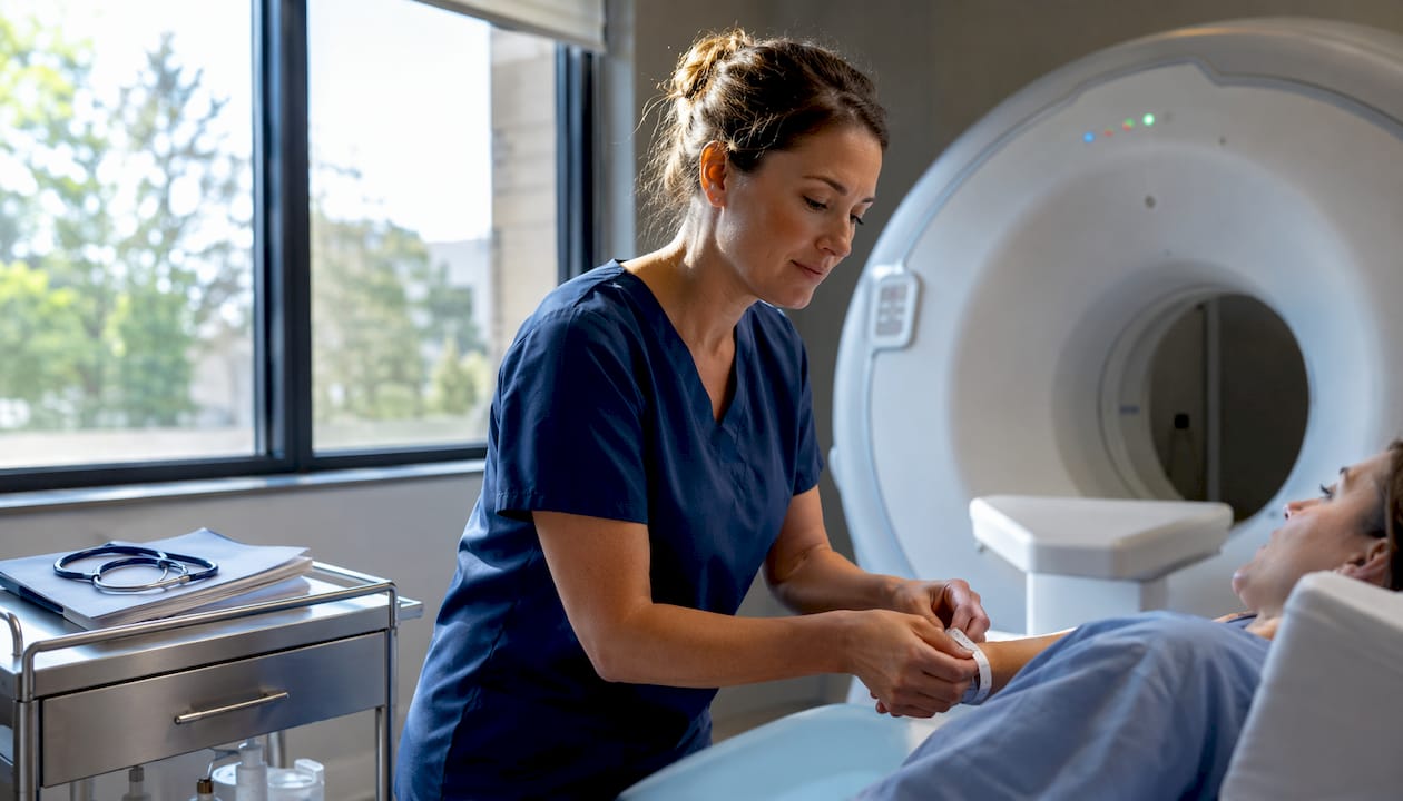
Most concussions do not require a brain scan. A brain scan for concussion is not a diagnostic test that confirms the injury — it is a safety screen designed to rule out life-threatening emergencies like bleeding or skull fracture. The American College of Radiology recommends CT imaging in the acute setting primarily for moderate-to-severe head trauma, not for every bump or collision. If you have no red-flag symptoms, then the more important next step is a specialist functional assessment covering vestibular, ocular, gait, and cognitive function.
When to seek emergency imaging immediately:
If none of these apply, your scan may well come back normal — and that is actually the expected result for most concussions, not a sign that something was missed.
The decision to image is driven by clinical red flags, not by the injury itself. In U.S. emergency departments, clinicians use validated decision tools — the Canadian CT Head Rule, NEXUS-II, and PECARN (for children) — to identify which patients genuinely need a CT scan and which can be safely observed without radiation exposure.
Practical timing guidance:
Pro Tip: If you are unsure whether your symptoms warrant an ER visit, err on the side of going. A clinician can apply a validated decision tool in minutes and spare you unnecessary radiation if imaging is not needed.
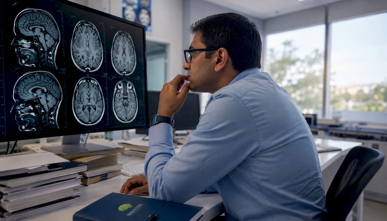
The Radiopaedia TBI overview confirms this split: CT is the first-line tool for acute hemorrhage and skull fracture, while MRI is preferred in the subacute or chronic phase when symptoms persist or CT findings are inconclusive.
The two modalities serve different purposes, and understanding the difference helps you ask better questions at your appointment.
| Feature | CT Scan | MRI |
|---|---|---|
| Acute bleed detection | Excellent | Good (SWI sequence) |
| Skull fracture | Excellent | Poor |
| Small contusion | Limited | Good |
| Diffuse axonal injury | Poor | Good (DWI, SWI) |
| Chronic sequelae | Limited | Excellent |
| Time to scan | a few minutes | over half an hour |
| Radiation | Yes (ionizing) | None |
| Availability/cost | Widely available, lower cost | Less available, higher cost |
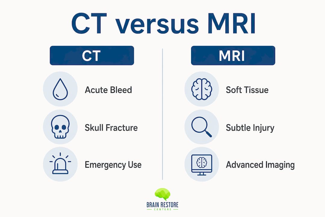
CT is fast and highly sensitive for hemorrhage, making it the workhorse for ER triage. MRI takes longer and costs more, but its specialized sequences add meaningful clinical detail. Diffusion-weighted imaging (DWI) can detect non-hemorrhagic axonal injury; susceptibility-weighted imaging (SWI) is more sensitive to microhemorrhages than standard CT; and FLAIR highlights edema and white-matter changes that CT simply misses.
When clinicians choose MRI over CT:
For sports-related concussion specifically, UMass Memorial Health notes that routine neuroimaging is reserved for suspected severe injury — most athletes with uncomplicated concussions never undergo imaging at all.
This is the part that confuses — and sometimes frustrates — patients most. A normal CT or MRI does not mean your concussion is not real or that your symptoms are imaginary.
“Concussion may result in neuropathological changes, but the acute clinical symptoms largely reflect a functional disturbance rather than a structural injury — and as such, no abnormality is seen on standard structural neuroimaging studies.” — 2012 Zurich Consensus Statement on Concussion in Sport
Concussion is primarily a metabolic and functional injury. The disruption happens at the cellular and microscopic level: ion channel dysfunction, altered neurotransmitter release, reduced cerebral blood flow. None of that shows up on a standard CT or MRI. The Queensland Brain Institute puts it plainly: visualizing concussion-related changes is challenging precisely because there is no obvious structural damage in most cases.
What this means practically:
Beyond standard CT and MRI, a growing set of advanced neuroimaging techniques can detect the microscopic and physiological changes that conventional scans miss. Most remain research tools, but a few are beginning to appear in specialty clinical settings.
Pro Tip: If a specialist recommends advanced imaging for persistent unexplained symptoms, ask whether the center uses standardized acquisition protocols and has normative comparison data — interpretation quality varies significantly between facilities.
The PMC review on TBI imaging notes that with wider adoption of conventional MRI, advanced techniques like DTI, MR perfusion, and MRS will increasingly guide care — but large longitudinal studies are still needed before they become routine diagnostic standards.
Imaging findings, or the absence of them, directly influence what happens next in your care.
Most people with concussion recover without any structural abnormality on imaging. When imaging is negative and symptoms continue, that is not a dead end — it is a clear signal to move toward vestibular, ocular, gait, and neurocognitive assessments that actually map how your brain is functioning.
During the scan:
Risks to know:
Insurance and cost realities:
Pro Tip: Ask for a copy of your DICOM files and the written radiology report before leaving. Bringing these to a concussion specialist or a post-accident clinic visit saves time and prevents repeat imaging.
At Brainrestoremeridian in Meridian, Idaho, imaging is the starting point, not the finish line. The clinic’s approach recognizes that most patients arrive with normal or near-normal scans and still need a clear path forward.
The clinic’s patient pathway:
Diagnostic and restorative tools available at the clinic:
If your symptoms have persisted beyond the typical recovery window, a concussion recovery checklist can help you organize your history before your first appointment.
Imaging rules out emergencies; functional assessment guides recovery — and most concussions require both in sequence, not imaging alone.
| Point | Details |
|---|---|
| CT for acute emergencies | Use CT within 24–48 hours when red-flag symptoms suggest bleeding or fracture. |
| MRI for persistent symptoms | Prefer MRI after 48 hours when CT is negative but neurological signs continue. |
| Normal scans are common | Most concussions are functional injuries; a normal scan does not invalidate symptoms. |
| Advanced imaging is largely research-stage | DTI, ASL, and fMRI show promise but lack standardized protocols for routine clinical use. |
| Brainrestoremeridian’s approach | The clinic combines imaging review with vestibular, ocular, and neurocognitive testing to build personalized recovery plans in Meridian, Idaho. |
The most common mistake I see patients make is treating a normal scan as a verdict. It is not. A clean CT or MRI tells you that your brain is not bleeding and your skull is intact — both genuinely important things to know. What it cannot tell you is why your concentration has dropped, why light bothers you, or why your balance feels off three weeks after the injury.
Functional assessments — vestibular testing, ocular motor evaluation, neurocognitive screening — answer those questions. Imaging and functional testing are not competing approaches; they are sequential ones. Get the emergency screen when the clinical picture calls for it, then move to the functional workup that actually maps your recovery. A normal scan paired with persistent symptoms is not a mystery. It is a clear signal that the next step is a multidisciplinary clinic, not another structural scan.
When imaging comes back normal but you still feel off, that is exactly the situation Brainrestoremeridian is built for. The clinic’s comprehensive brain health restoration program in Meridian, Idaho integrates neurofeedback, photobiomodulation, vestibular and ocular rehabilitation, and functional medicine into a single coordinated plan — built around your imaging results and functional test findings, not a one-size-fits-all protocol.
Bring your ER records, any prior imaging (DICOM files and radiology reports), and a written symptom log to your first visit. The team uses that information alongside hands-on functional testing to identify exactly where your recovery has stalled. To schedule a consultation and start building your personalized plan, visit Brainrestoremeridian.
This article is general health information, not medical advice. Confirm your specific situation with a qualified clinician or primary care provider.
Research and academic sources:
Clinic resources (Brainrestoremeridian):
Questions about your next step?
Our Meridian team can help you understand the evaluation process and which care options may fit your needs.
Schedule a Brain Health ConsultationPrefer to speak with our team? Call 208-639-1397