Stress Shrinks Hippocampus: The Science and How to Recover
August 19, 2026

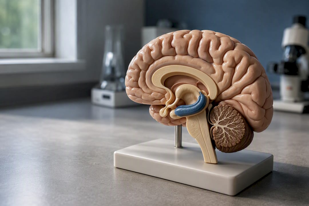
Yes, chronic stress can shrink the hippocampus. It happens because prolonged activation of your stress response floods this memory center with cortisol, and that excess hormone causes hippocampal neurons to retract their dendrites while slowing the birth of new brain cells. The good news sits alongside the bad news: most of this “shrinkage” reflects flexible, living tissue pulling back, not neurons dying off permanently. Recovery is possible for many people, though how much and how fast depends on how long the stress lasted and how severe it was.
Here’s the short version of what’s happening inside your brain when chronic stress takes hold:
Cortisol and cognition are closely linked. In Cushing’s syndrome, a condition marked by chronically elevated cortisol, 83% of patients report memory impairment and 66% report a shortened attention span. That single data point tells you almost everything you need to know about why stress and memory loss travel together.
Chronic stress shrinks the hippocampus mainly through dendritic retraction and reduced neurogenesis driven by sustained cortisol exposure, and this process is often reversible with the right intervention.
| Point | Details |
|---|---|
| Shrinkage is usually structural, not fatal | Dendrites retract and spines shrink, but neurons typically survive and can regrow. |
| Cortisol drives the damage | Chronic HPA axis activation keeps glucocorticoids elevated, overstimulating hippocampal receptors. |
| Cognitive symptoms are common | In Cushing’s syndrome, a large majority of patients report memory impairment and many report shortened attention span. |
| Recovery depends on duration and severity | Shorter stress exposure generally predicts more complete recovery once the stressor ends. |
| Multidisciplinary support helps | Brainrestoremeridian combines neurofeedback, photobiomodulation, and functional medicine assessment to support recovery. |
The hippocampus sits at the intersection of memory, spatial navigation, and emotional regulation, which makes it unusually sensitive to hormonal traffic. It’s also packed with glucocorticoid receptors, more than almost any other brain region, so when cortisol rises and stays elevated, this structure absorbs the impact first and hardest.
Understanding why stress affects memory requires separating two different bodies of evidence: what scientists can prove by manipulating stress directly in animals, and what they can only observe by association in humans.
Animal research established causation decades ago. Researchers exposing rodents to repeated restraint stress or sustained corticosterone (the rodent equivalent of cortisol) consistently observe dendritic debranching in the CA3 subregion and reduced neurogenesis in the dentate gyrus. These structural changes track closely with worse performance on hippocampal-dependent tasks like maze navigation, giving scientists a direct causal link between stress hormone exposure and both brain structure and behavior.
Human imaging work tells a complementary, if messier, story. A large-scale analysis using the UK Biobank cohort found that lifetime stress exposure correlates with changes in limbic microstructure along with worse executive function and working memory, with effects that differ somewhat by sex and by the life stage when stress occurred. Clinical populations add another layer: people with Cushing’s syndrome, whose bodies overproduce cortisol for months or years, show smaller hippocampal and cortical volumes that correlate directly with how long they’ve had the disease. Patients with PTSD and major depression show similar, if more variable, patterns on MRI.
The hippocampus is one of the most plastic structures in the adult brain. It shrinks under sustained glucocorticoid pressure, but that same plasticity is what makes recovery possible once the pressure lifts.
Here’s where the evidence gets nuanced rather than simple:
Taken together, the animal work explains the mechanism and the human work confirms it plays out in real lives, even if the human picture carries more static.
The path from a stressful life to a smaller-looking hippocampus on a brain scan runs through a fairly specific chain of biology, and knowing the steps matters because each one represents a point where intervention might help.
It starts with a stressor, real or perceived, that activates your HPA axis. Your hypothalamus releases corticotropin-releasing hormone (CRH), which prompts your pituitary gland to release ACTH, which signals your adrenal glands to pump out cortisol. In a healthy stress response, cortisol does its job and levels drop back down within hours. Under chronic stress, that shutoff mechanism weakens and cortisol stays elevated for weeks, months, or years.
Once cortisol is chronically high, it binds to two receptor types in the hippocampus: mineralocorticoid receptors (MR) and glucocorticoid receptors (GR). At normal cortisol levels, these receptors support healthy synaptic function. Under sustained high cortisol, GR activation shifts the balance and sets off a cascade involving excess glutamate release and overactivation of NMDA receptors. That excitatory overload changes the cell’s internal skeleton, and dendrites, the branch-like structures neurons use to receive signals from neighboring cells, begin retracting.
The principal molecular players in this cascade include:
At the same time, neurogenesis in the dentate gyrus, the ongoing production of new neurons that continues throughout adulthood, slows down. Elevated glucocorticoids suppress the stem cell activity that would otherwise generate fresh neurons, and lower BDNF removes some of the growth signal those new cells need to survive and integrate.
Pro Tip: The distinction between dendritic retraction and actual neuron death matters enormously for what you should expect from treatment. Retraction is a structural pullback, similar to a tree shedding branches in a hard winter. The root system, the neuron itself, is usually still alive. That’s very different from neuronal death, where the cell itself is gone. Most stress-related hippocampal changes fall into the retraction category, which is why recovery is on the table for so many people.
If you wanted to sketch this as a diagram, it would run left to right: stressor → HPA axis activation → chronic cortisol elevation → MR/GR receptor overactivation → glutamate/NMDA excitotoxic signaling → dendritic retraction and suppressed neurogenesis → measurable volume loss on MRI. Research on this pathway also shows stress effects aren’t confined to the hippocampus; while hippocampal dendrites shrink under chronic stress, dendrites in the basolateral amygdala often expand, which helps explain why chronic stress can leave you more reactive to threats while your reflective, contextual memory gets weaker at the same time.
When you read that stress “shrinks” the hippocampus, that claim usually rests on one specific technique: MRI volumetry. Researchers take structural brain scans, trace the boundaries of the hippocampus using standardized atlases, and calculate its total volume in cubic millimeters. Compare that volume across groups, or across time in the same person, and you get a number you can analyze statistically.
That method is powerful, but it measures something coarser than what’s actually happening at the cellular level. MRI volumetry captures macrostructure, the overall size of the region. It cannot see individual dendrites shrinking or new neurons being born. Animal studies fill that gap using histology, physically examining brain tissue under a microscope to count dendritic branch points, spine density, and newly formed cells. That’s a big part of why animal and human data feel like they’re speaking different dialects of the same language: one measures the forest, the other counts individual branches.
Several methodological issues complicate how you should read human MRI findings:
Group-level differences in these studies, meanwhile, are statistical averages. A study might find that a chronically stressed group has, on average, smaller hippocampal volume than a control group. That doesn’t mean every stressed individual in that group shows shrinkage, and it doesn’t mean your personal risk can be read off a population trend. Individual variation is large, which is exactly why a personalized clinical assessment matters more than a headline statistic ever could.
Structural change in the hippocampus doesn’t stay abstract for long. It shows up in daily life as specific, recognizable difficulties, and knowing what to watch for helps you connect the dots between “I’ve been under a lot of stress” and “why can’t I remember where I put my keys anymore.”
The hippocampal-dependent functions most commonly affected include:
These aren’t minor inconveniences. Struggling to hold onto new information at work, forgetting commitments to family, or losing your sense of direction in familiar places all trace back to the same hippocampal circuitry under strain.
The mood connection runs both directions. Chronic stress and hippocampal changes are closely tied to depression and PTSD, and the relationship is bidirectional. Reduced hippocampal volume shows up as a common finding in both conditions, and hippocampal dysfunction itself may impair the ability to regulate emotional responses, since the region normally helps put fear and threat memories into proper context. Fewer hippocampal brakes on the amygdala’s alarm system can mean more anxiety, more intrusive memories, and a harder time feeling safe even when nothing dangerous is present.
None of this plays out identically for everyone. Effect sizes vary substantially across individuals, and some people show remarkable resilience despite significant stress exposure, while others show measurable changes after what looks like a moderate stressor. Genetics, social support, and coping style all shape where someone lands on that spectrum, which is a major reason two people can go through similar circumstances and come out with very different cognitive outcomes.
This is the question most people actually want answered, and the honest answer is: often, yes, though the details depend heavily on how long and how severe the stress was.
Animal research offers the clearest timeline evidence. When researchers remove a chronic stressor from rodents, dendritic regrowth in CA3 neurons and functional recovery on memory tasks can occur within weeks. The same plasticity that made the hippocampus vulnerable to shrinkage also makes it capable of rebuilding once the hormonal pressure lifts.
Human evidence, while less precise on timing, points in the same direction. Patients who undergo successful surgical treatment for Cushing’s syndrome, removing the source of chronic cortisol overproduction, show partial recovery of hippocampal volume on follow-up MRI scans, along with improvement in memory symptoms. Recovery tends to track with how long the hypercortisolemia lasted before treatment; shorter disease duration generally predicts more complete recovery.
The biological mechanisms that drive this rebuilding process include:
Real gaps remain in this picture, and it’s worth being upfront about them:
What the evidence does support is a straightforward principle: removing the chronic stressor and giving the brain time and the right physiological conditions gives it the best chance to rebuild. That’s a fundamentally more hopeful message than the phrase “brain shrinkage” tends to suggest on its own.
Not everyone facing chronic stress ends up with the same degree of hippocampal change, and several factors shift both risk and recovery potential in predictable ways.
If you’re noticing persistent memory decline, difficulty concentrating that’s interfering with work or relationships, or emotional symptoms that aren’t improving, that’s a signal worth taking to a clinician rather than working around indefinitely. Early evaluation gives you more options and generally better outcomes than waiting until symptoms become entrenched.
The research on stress-induced hippocampal changes points toward a handful of interventions with real evidence behind them, and prioritizing matters more than trying to do everything at once.
On timing, don’t expect overnight results. Animal studies show dendritic regrowth within weeks once a stressor is removed, but human cognitive improvement tends to unfold over months rather than weeks, particularly if the stress was long-standing. Structural recovery on imaging, where it’s been documented, typically lags behind subjective symptom improvement.
Pro Tip: Don’t try to overhaul everything at once. Pick one or two high-impact changes, sleep and exercise are the strongest combination, and track them consistently for a month before adding more. Trying to fix your diet, sleep, exercise, and therapy schedule simultaneously usually backfires because none of it sticks.

If you’re dealing with persistent cognitive symptoms, a clinical evaluation typically includes neuropsychological testing to map which specific memory and attention functions are affected, sometimes structural MRI to assess hippocampal volume directly, and an endocrine screen to check for elevated cortisol or an underlying condition like Cushing’s syndrome. Support for poor focus and concentration often starts with exactly this kind of assessment rather than guesswork.
Beyond lifestyle changes, a range of clinical and device-based approaches are used to support brain recovery after chronic stress, each with a different evidence profile worth understanding honestly before you commit to one.
Neurofeedback trains the brain toward healthier electrical activity patterns using real-time EEG feedback, and it’s increasingly used to support attention and emotional regulation in people recovering from chronic stress or anxiety. Evidence strength: promising, with a growing but still developing research base, particularly for anxiety-related symptoms where neurofeedback shows measurable benefit.
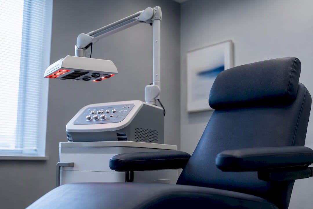
Photobiomodulation (laser therapy) uses specific wavelengths of light believed to support cellular energy production in neurons, with some clinical use in neurological stress and recovery contexts. Evidence strength: preliminary to promising, an active area of research rather than an established standard.
Hyperbaric oxygen therapy increases oxygen delivery to tissue under pressure, with proposed benefits for neurological recovery and inflammation reduction. Evidence strength: preliminary for stress-related cognitive symptoms specifically, though better established for certain other medical indications.
Functional medicine strategies look at the whole physiological picture, hormone balance, inflammation, gut health, sleep architecture, to identify factors that may be perpetuating chronic stress physiology. Evidence strength: variable by specific intervention, and best used as a framework for personalized assessment rather than a single standardized treatment.
If you’re evaluating providers offering these therapies, look for practitioners who combine multiple approaches rather than promising one modality will fix everything, ask direct questions about what outcomes to realistically expect and by when, and confirm any device-based therapy has appropriate safety screening for your specific health history.
| Modality | Evidence Strength | Best Used For |
|---|---|---|
| Neurofeedback | Promising | Attention and emotional regulation support |
| Photobiomodulation | Preliminary to promising | Cellular-level neurological support |
| Hyperbaric oxygen therapy | Preliminary (for stress-related cognition) | Oxygenation and inflammation support |
| Functional medicine assessment | Variable, framework-dependent | Identifying perpetuating physiological factors |
Most people recovering from stress-related cognitive changes do best with multidisciplinary care, combining behavioral changes, medical evaluation, and where appropriate, device-based or functional medicine support, rather than betting everything on a single therapy.
Knowing the actual biology behind hippocampal shrinkage changes how you think about recovery, and I’d argue that matters more than most people realize. When “stress shrinks your brain” gets flattened into a scary headline, it sounds like permanent damage. When you understand it’s largely dendritic retraction, a plastic, responsive process rather than cell death, the whole conversation shifts from fear to strategy.
That distinction isn’t just academic reassurance. It changes what you should actually do. If you believed the damage was fixed and irreversible, you might reasonably give up on lifestyle changes or treatment. Once you understand the hippocampus is built to rebuild given the right conditions, sleep, reduced cortisol load, exercise, targeted therapy, the entire recovery conversation becomes about creating those conditions rather than accepting a diagnosis as a life sentence.
I’d also push back gently on the idea that recovery is purely a matter of willpower or stress management alone. For some people, the physiological damage from years of elevated cortisol needs more than meditation and better sleep hygiene; it needs an actual clinical evaluation. If persistent memory problems, mood changes, or cognitive fog are interfering with your work or relationships, that’s the signal to get assessed rather than push through it indefinitely.
Brainrestoremeridian is built specifically around the science covered here: helping you address the physiological roots of stress-related cognitive change, not just manage symptoms around the edges. Our multidisciplinary approach combines neurofeedback, photobiomodulation, functional medicine assessment, and structured cognitive evaluation, all under one roof in Meridian, Idaho, so you’re not piecing together a recovery plan from disconnected providers.

A first visit typically includes a thorough intake covering your stress history and symptoms, cognitive screening to identify which specific functions (memory, attention, processing speed) are affected, and a personalized care plan drawing from the modalities that fit your situation. If you’re wondering how operant conditioning principles shape neurofeedback training, that evaluation is where you’ll get a clear, individualized answer rather than a generic protocol.
This isn’t a substitute for emergency care. If you’re experiencing sudden, severe cognitive changes or a medical crisis, seek immediate medical attention. For persistent stress-related memory and focus concerns, schedule an evaluation to start mapping out what recovery looks like for you specifically.
Does stress permanently shrink the hippocampus?
Usually not. Most stress-related hippocampal shrinkage reflects dendritic retraction and reduced neurogenesis, both plastic processes that can reverse once chronic stress ends, though recovery depends on how long and severe the stress was.
How long does it take for the hippocampus to recover after chronic stress?
Animal studies show measurable dendritic regrowth within weeks of removing a chronic stressor. Human recovery timelines are less precise but generally unfold over months, especially after long-standing stress.
Can exercise reverse hippocampal shrinkage?
Regular aerobic exercise supports neurogenesis and raises BDNF levels, both of which favor hippocampal recovery, making it one of the most consistently supported lifestyle interventions available.
What are the signs that stress has affected your hippocampus?
Common signs include difficulty forming new memories, trouble navigating familiar places, struggling to hold context around events, and mood changes like increased anxiety or depressive symptoms.
Is hippocampal shrinkage the same as brain damage?
Not typically. It’s more accurate to describe it as structural remodeling, reduced dendritic complexity rather than widespread neuron death, which is why the outlook for recovery is generally more hopeful than the word “shrinkage” suggests.
This article is general information, not a substitute for advice from a qualified doctor. Consult a qualified healthcare professional about your own circumstances before acting on anything here.
Consult a clinician for personalized interpretation of any imaging results or persistent symptoms; population-level research can’t substitute for an individual assessment.
Questions about your next step?
Our Meridian team can help you understand the evaluation process and which care options may fit your needs.
Schedule a ConsultationPrefer to speak with our team? Call 208-639-1397