Acetylcholine and Memory Diseases: A Clinical Guide
August 16, 2026

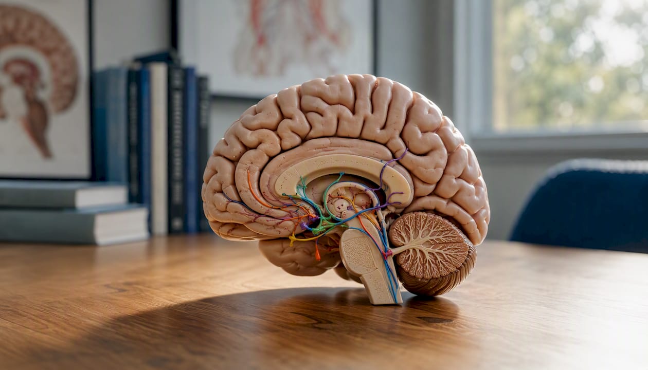
Acetylcholine (ACh) is a central, dynamic modulator of memory whose loss contributes directly to cognitive decline in diseases such as Alzheimer’s, Lewy body dementia, and vascular cognitive impairment. Postmortem studies, neuroimaging, and decades of clinical trials all point to the same conclusion: cholinergic signaling is indispensable for encoding new memories and sustaining attention, and its progressive failure tracks closely with the severity of memory loss. Approved acetylcholinesterase (AChE) inhibitors offer real, measurable symptomatic benefit, but they do not slow or reverse the underlying disease process. Understanding the role of acetylcholine in memory diseases matters because it shapes realistic expectations, guides clinical conversations, and points toward where the next generation of therapies must go. This article is for informational purposes only and is not a substitute for individualized medical evaluation or treatment by a qualified clinician.
Key points at a glance:
Acetylcholine is a dynamic memory modulator whose loss in Alzheimer’s disease tracks directly with cognitive severity, and while AChE inhibitors offer real symptomatic benefit, no approved cholinergic therapy has yet demonstrated disease modification.
| Point | Details |
|---|---|
| Cholinergic loss drives memory decline | Nucleus basalis of Meynert neuron counts fall from roughly 500,000 to fewer than 100,000 in advanced Alzheimer’s disease. |
| AChE inhibitors are symptomatic only | Donepezil, galantamine, and rivastigmine improve cognition modestly but do not slow underlying neurodegeneration. |
| Bidirectional ACh regulation matters | Both deficiency and excess of central ACh can impair learning; dose titration must be gradual and individualized. |
| Receptor subtypes shape therapeutic targets | M1 muscarinic and α7 nicotinic receptors are the most promising targets for encoding-specific, side-effect-reduced future drugs. |
| Multimodal care extends beyond ACh | Non-pharmacologic approaches that support brain network function can complement cholinergic therapies in a comprehensive care plan. |
The cholinergic system does not simply “turn memory on.” ACh acts as a gain-control signal, raising the brain’s sensitivity to incoming information during active encoding while suppressing the feedback loops that would otherwise replay stored patterns and interfere with learning. When that signal degrades, as it does early and severely in Alzheimer’s disease, the brain loses its ability to write new memories even as older ones remain relatively accessible for a time.
Cholinergic modulation of the hippocampal region is central to this process. Experimental work consistently links ACh signaling to hippocampal synaptic plasticity and the attention-related processing that primes circuits for encoding. This is not a peripheral finding. It is the mechanistic foundation on which every approved memory drug is built.
The central cholinergic system is organized around two main groups of projection neurons. Understanding their anatomy explains why memory is so vulnerable when they degenerate.
Basal forebrain cholinergic nuclei are the primary source of ACh for the cortex and hippocampus. The key structures include:
These nuclei project to virtually every region involved in memory consolidation: hippocampus, prefrontal cortex, parietal association cortex, and amygdala. Their output regulates cortical state, attention, and the signal-to-noise ratio for incoming sensory information.
Beyond neurons, glial cells express cholinergic receptors and respond to ACh release. Astrocytes, for instance, can modulate synaptic ACh clearance and contribute to the local cholinergic microenvironment, though this non-neuronal signaling is less well characterized than neuronal projections.
Statistic to know: In advanced Alzheimer’s disease, nucleus basalis of Meynert neuron counts fall from roughly 500,000 in healthy adults to fewer than 100,000, a loss of more than 80% of the primary cortical ACh supply.
ACh acts on two broad receptor families, and their distinct properties explain why the same neurotransmitter can produce such varied effects depending on where and when it is released.
Muscarinic receptors are G-protein-coupled and therefore slower-acting. Five subtypes exist (M1–M5). M1 receptors dominate in the cortex and hippocampus and are postsynaptic; they enhance excitability, support persistent spiking in pyramidal neurons, and facilitate long-term potentiation (LTP). M2 receptors sit presynaptically on cholinergic terminals and act as autoreceptors, limiting ACh release. M4 receptors modulate dopaminergic circuits relevant to attention.
Pharmacological blockade of muscarinic receptors selectively impairs encoding of new information while leaving retrieval of previously stored memories relatively intact. This dissociation is one of the clearest pieces of evidence that muscarinic signaling is specifically required for writing new memory traces, not reading old ones.
Nicotinic receptors are ligand-gated ion channels and respond in milliseconds. The α7 subtype is highly expressed in the hippocampus and prefrontal cortex; it has high calcium permeability and is closely linked to LTP induction and attention. The α4β2 subtype modulates thalamo-cortical gating and is a target of several investigational drugs.
| Feature | Muscarinic (mAChR) | Nicotinic (nAChR) |
|---|---|---|
| Receptor type | G-protein-coupled (metabotropic) | Ligand-gated ion channel (ionotropic) |
| Response speed | Slow (seconds) | Fast (milliseconds) |
| Key subtypes for memory | M1 (postsynaptic), M2 (presynaptic autoreceptor) | α7, α4β2 |
| Primary memory role | Encoding, persistent spiking, LTP facilitation | Attention, LTP induction, thalamo-cortical gating |
| Effect on glutamate | Modulates NMDA receptor sensitivity | Enhances glutamate release presynaptically |
| Pharmacologic targets | M1 agonists, M2 antagonists, PAMs | α7 agonists, α4β2 partial agonists |
Both receptor families modulate glutamatergic transmission. M1 activation increases NMDA receptor sensitivity, which lowers the threshold for LTP. α7 nAChRs on glutamatergic terminals facilitate vesicle release, amplifying excitatory drive during periods of high cholinergic tone. The net result is that ACh, when present at the right level and time, primes hippocampal circuits for plasticity.
Think of ACh as a switch between two operating modes. At high levels, the brain favors encoding: it amplifies incoming sensory signals and suppresses the recurrent feedback connections that would otherwise dominate processing. At low levels, the balance tips toward retrieval, allowing stored patterns to be reactivated.
This encoding-retrieval shift has strong experimental support:
The timing of cholinergic signaling relative to learning events is essential: ACh favors encoding if present during afferent-driven activity but may interfere with retrieval if elevated at the wrong moment. This is not a theoretical concern. It has direct implications for how AChE inhibitors are dosed and for why simply flooding the synapse with ACh is not always beneficial.
Muscarinic receptor blockade studies confirm the encoding specificity: scopolamine, a muscarinic antagonist, reliably impairs new learning in healthy volunteers while leaving recall of pre-drug information largely unaffected.
The cholinergic hypothesis of Alzheimer’s disease, first articulated in the early 1980s, has accumulated substantial supporting evidence across multiple lines of inquiry.
Postmortem and neuroimaging findings:
Clinical and epidemiologic correlations:
Mechanistic links to amyloid and tau:
AChE itself can interact with amyloid precursor protein processing and amyloid-beta aggregation, creating a bidirectional relationship between cholinergic enzymes and amyloid biology. Reduced ACh signaling may also promote tau hyperphosphorylation and neuroinflammation, meaning that cholinergic failure is not simply a downstream consequence of amyloid pathology but an active contributor to it.
Beyond Alzheimer’s disease:
Cholinergic involvement is not unique to Alzheimer’s. In Lewy body dementia, cholinergic deficits are often more severe than in Alzheimer’s disease at equivalent stages, which may explain why patients with Lewy body dementia sometimes show a more pronounced response to AChE inhibitors. Vascular cognitive impairment disrupts cholinergic white-matter pathways connecting the basal forebrain to cortical targets, producing a pattern of attentional and executive dysfunction that overlaps with, but differs from, the encoding-dominant deficit of Alzheimer’s disease.
A systems-level review positions Alzheimer’s as a multifactorial disease in which cholinergic failure is an early and important contributor but interacts with glutamatergic, GABAergic, monoaminergic, inflammatory, and vascular dysfunction. That framing matters for treatment: single-target cholinergic drugs address one node in a much larger network.
Three AChE inhibitors are approved for Alzheimer’s disease in the United States. All three work by slowing the enzymatic breakdown of ACh in the synapse, raising the concentration of available transmitter. Their differences lie in selectivity, additional mechanisms, and tolerability profiles.
Donepezil is a selective, reversible AChE inhibitor approved for all stages of Alzheimer’s disease, from mild to severe. It is typically taken once daily, which supports adherence. Common side effects include nausea, diarrhea, insomnia, and vivid dreams, most of which are dose-dependent and tend to diminish over time. Donepezil produces consistent, modest improvements on standardized cognitive scales, particularly in attention and verbal memory domains.
Rivastigmine inhibits both AChE and butyrylcholinesterase (BuChE), a second cholinergic enzyme whose activity increases as AChE declines in Alzheimer’s disease. This dual inhibition may provide broader cholinergic coverage. Rivastigmine is available as an oral formulation and a transdermal patch; the patch significantly reduces gastrointestinal side effects compared to oral dosing. It is approved for mild-to-moderate Alzheimer’s disease and for Parkinson’s disease dementia, where cholinergic deficits are also prominent.
Galantamine inhibits AChE and also acts as a positive allosteric modulator (PAM) of nicotinic receptors, amplifying the response to endogenous ACh at nAChRs. This dual action is pharmacologically distinct from the other two agents. Galantamine is approved for mild-to-moderate Alzheimer’s disease. Gastrointestinal side effects are the most common tolerability concern, and extended-release formulations improve this profile.
On evidence strength: Clinical-trial data consistently show symptomatic cognitive improvements with AChE inhibitors, but effect sizes are modest and no trial has demonstrated disease modification. These drugs slow the functional expression of decline in some patients; they do not halt or reverse the underlying neurodegeneration. Setting that expectation clearly with patients and caregivers is one of the most important things a clinician can do.
A critical dosing caution: ACh has a bidirectional relationship with memory. Physiologic increases can enhance encoding, but excessive central ACh can impair learning. Aggressive dose escalation of AChE inhibitors does not linearly improve cognition and may worsen it in some patients, particularly those with preserved cholinergic tone or concurrent use of other cholinergic agents. Dose titration should be gradual and individualized.
The limitations of nonselective AChE inhibition have pushed research in several directions, each addressing a different gap in the current therapeutic picture.
Bidirectional regulation and dosing precision:
The finding that both ACh deficiency and excess can impair learning is not just a pharmacological curiosity. It suggests that future therapies need to restore physiologic cholinergic tone, not simply maximize it. Receptor-subtype-selective drugs and positive allosteric modulators are designed with this in mind.
Receptor-selective strategies:
Biomarker development:
Measuring cholinergic integrity in living patients remains a significant challenge. PET ligands targeting vesicular acetylcholine transporter (VAChT) and other cholinergic markers are under active development, but current tracers have limited sensitivity and have not yet translated to routine clinical use. Peripheral confounds, including cholinergic activity in blood and muscle, complicate interpretation of systemic markers.
Neuroprotective and combination approaches:
Trophic-factor strategies, particularly nerve growth factor (NGF) delivery to basal forebrain neurons, aim to preserve cholinergic neurons rather than compensate for their loss. Early gene-therapy trials have shown proof-of-concept but face delivery challenges. Combination approaches that pair cholinergic support with anti-amyloid, anti-tau, or anti-inflammatory interventions reflect the systems-level understanding that no single target is sufficient.
The translational gap:
For disease-modifying cholinergic therapies to succeed, trials need to enroll patients earlier, use cholinergic biomarkers as enrollment and outcome criteria, and design endpoints that capture encoding-specific cognitive domains rather than global scales that are insensitive to early change. That infrastructure is still being built.
Understanding the science is one thing. Knowing what to do with it is another. Here is what the evidence translates to in practical terms.
Realistic expectations for AChE inhibitors:
Monitoring and safety:
When to consider specialist referral or trial enrollment:
Non-pharmacologic approaches, including neurofeedback for neurodegenerative patients, can complement cholinergic therapies by supporting brain network regulation through mechanisms that do not depend on ACh levels alone. Exploring integrated strategies for cognitive decline alongside pharmacologic management gives you the broadest possible foundation for care.
Pro Tip: When meeting with a neurologist or memory specialist, ask specifically: “What cognitive domains should I expect this medication to help with, over what time frame, and how will we know if it’s working?” That question moves the conversation from generic reassurance to measurable, trackable goals.
The mechanistic picture of cholinergic failure in memory diseases is genuinely clarifying, but it also reveals how much the field has been working with an incomplete map. The cholinergic hypothesis gave us useful drugs and a productive research framework, yet the modest effect sizes of AChE inhibitors have always hinted that ACh is one critical node in a much larger, interconnected system.
What strikes me most about the current evidence is the bidirectional regulation finding. The instinct in clinical practice, and in drug development, has been to treat cholinergic deficiency as a simple deficit to be corrected by raising ACh. The data say otherwise. Timing, receptor subtype, and circuit context all determine whether more ACh helps or hurts. That complexity is not a reason for pessimism. It is a reason to move toward more precise, individualized approaches.
At Brainrestoremeridian, the cholinergic science informs how we think about assessment and care planning. Brain network function, as measured through tools like qEEG, reflects the downstream consequences of neurotransmitter dysregulation, including cholinergic dysfunction. Approaches like neurofeedback, photobiomodulation, and functional medicine strategies that address neurodegeneration do not replace pharmacologic treatment, but they address dimensions of brain health that AChE inhibitors do not touch. For patients navigating memory concerns, that multimodal perspective can make a real difference in how they function day to day. This perspective reflects general clinical reasoning and is not a substitute for individualized medical advice from your own clinician.

Questions about your next step?
Our Meridian team can help you understand the evaluation process and which care options may fit your needs.
Schedule a Brain Health ConsultationPrefer to speak with our team? Call 208-639-1397